


Bethlehem / PNN /
Palestinian ophthalmologist Dr. Omar Hammad has succeeded in modifying a standard German-made ophthalmic microscope into a three-dimensional system, a development that significantly facilitates his work as a surgeon and increases the success rates of eye operations through 3D imaging technology.
The achievement represents a victory of intellect and determination.
Dr. Hammad said he considers his work both a Palestinian and Arab achievement, expressing hope that it will contribute to advancing scientific research and supporting innovative initiatives. His locally developed 3D microscope competes with advanced German 3D microscopes that cost around $300,000, while his upgraded system costs only a few thousand dollars in addition to the original microscope price.
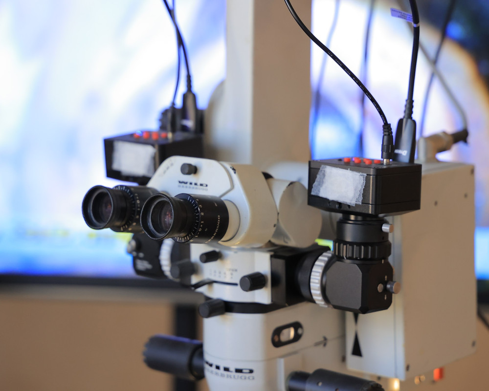
Explaining the details of the development, Dr. Hammad said the project involved adding cameras and a three-dimensional imaging system at a cost of only $20,000. Over the past year, he closely followed specialized scientific conferences and publications. The idea to develop the microscope emerged after he attended an international conference on the subject and asked a specialized professor about certain technical details of their 3D system, but the professor declined to answer.
Dr. Hammad said he conducted extensive studies comparing conventional microscopes with 3D microscopes, analyzing the differences between them before beginning work on upgrading his own device. He purchased components from manufacturing companies online and then developed a clear implementation plan.
Work in stages
According to Dr. Hammad, the plan was divided into two stages. The first focused on purchasing and installing equipment, including cameras, sensors, screens and 3D glasses. After installing and testing the components, he achieved improved vision that allowed him to see depth more clearly, enabling him to work with greater physical and psychological comfort as an eye surgeon.
After installing the cameras and sensors, Dr. Hammad worked on generating and integrating 3D images using a high-speed, high-performance computer system. This allowed him to connect two screens side by side, similar to those used in expensive commercial 3D microscopes. He then installed specialized 3D imaging software, achieving results that rival modern high-end devices.
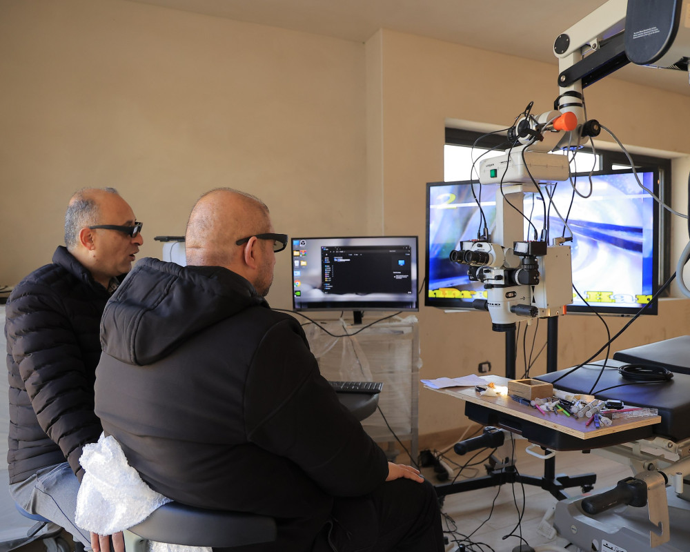
The second stage, which is currently underway, involves conducting experimental surgeries based on the results of the first stage. Dr. Hammad is performing procedures on animal eyes, including fisheyes. Initial results have been very positive, allowing him to operate with ease and contributing to higher success rates.
Technical obstacles overcome
Dr. Hammad said the project faced numerous challenges, beginning with the absence of institutional support for scientific research and development. Despite this, his determination pushed him to continue, covering all equipment costs from his own personal funds.
A second challenge involved sourcing the required components, as few companies were willing to sell directly to individuals. This forced him to spend long hours online communicating with manufacturing representatives. Although the process was difficult, his persistence enabled him to obtain the necessary equipment.
The third challenge related to integrating and synchronizing the technologies. He sought assistance from specialists, some of whom were skeptical of his work. Nevertheless, he overcame these obstacles through research, experimentation and problem-solving.
One of the most complex technical issues involved calculating image latency — the delay between image capture and its display on the screen. Commercial systems that address this issue were prohibitively expensive. Instead, Dr. Hammad devised a method using two mobile phones: one mounted under the camera and the other on the display screen, both activated simultaneously to measure the delay. He found that the latency was only fractions of a second, allowing him to accurately address and overcome the issue.
Proven success
Videos presented by Dr. Hammad demonstrate the success of the experimental procedures at all stages. Through 3D glasses, distinct layers of the eye can be seen with clear depth, separated smoothly and efficiently — confirming the success of the experiment.
Dr. Hammad said he now performs experimental surgical procedures with greater ease, as operating via a 3D screen provides enhanced control without physical strain. The 3D display system also allows nursing and medical staff assisting in the operation to closely follow the procedure.
He stressed that his goal in developing the microscope is not commercial gain, but rather to contribute to improving ophthalmic services in Palestine and across the Arab world. He noted that standard microscopes can be converted into 3D systems, a development with significant medical implications for both patients and surgeons.
Dr. Hammad added that once all experiments are completed, he plans to brief medical and media institutions in Palestine, particularly the Palestinian Ministry of Health. He expressed hope that the ministry will adopt the innovation and convert its microscopes to 3D systems based on his research and development success.
This story was produced as part of the Qarib project, implemented by the French media development agency CFI in partnership with and funded by the French Development Agency (AFD).